OVERVIEW
In the complexity of modern medicine, diagnosis and technology there is an increasing challenge to professionals to engage with patients that enable effective screening and lifestyle changes before clinical intervention.
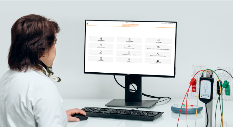
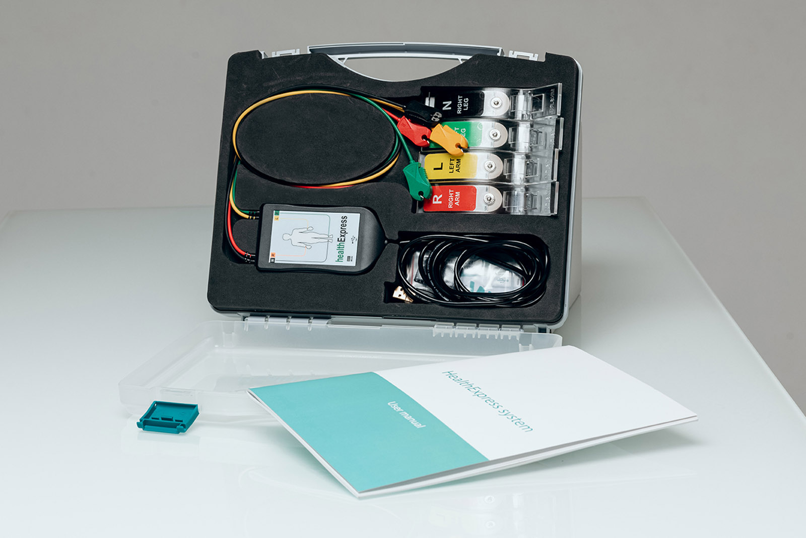
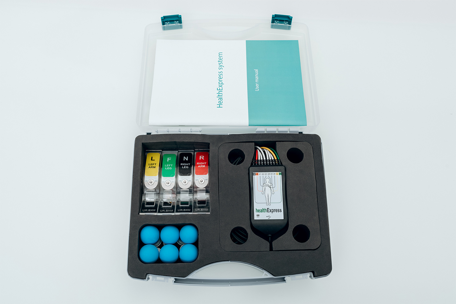
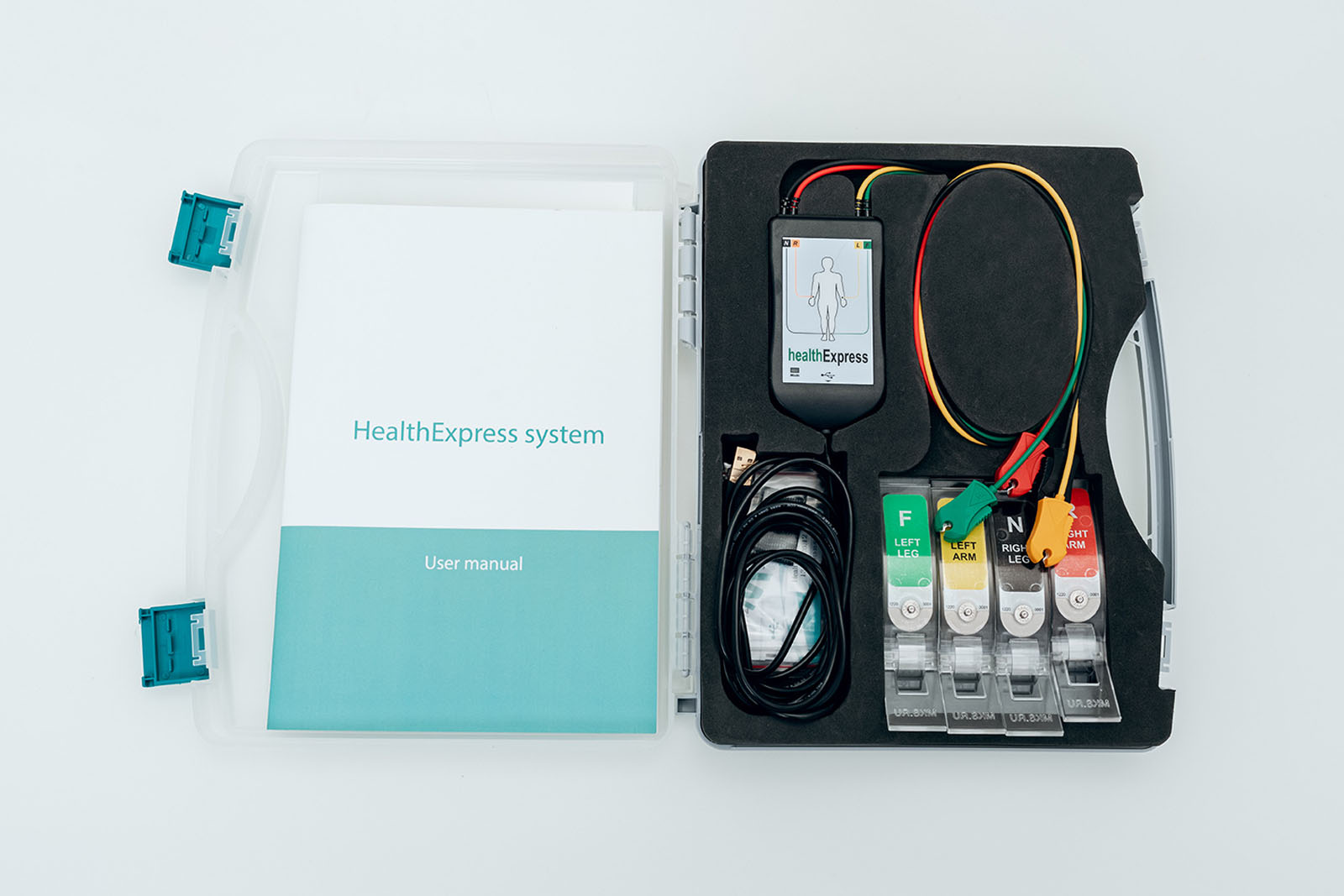
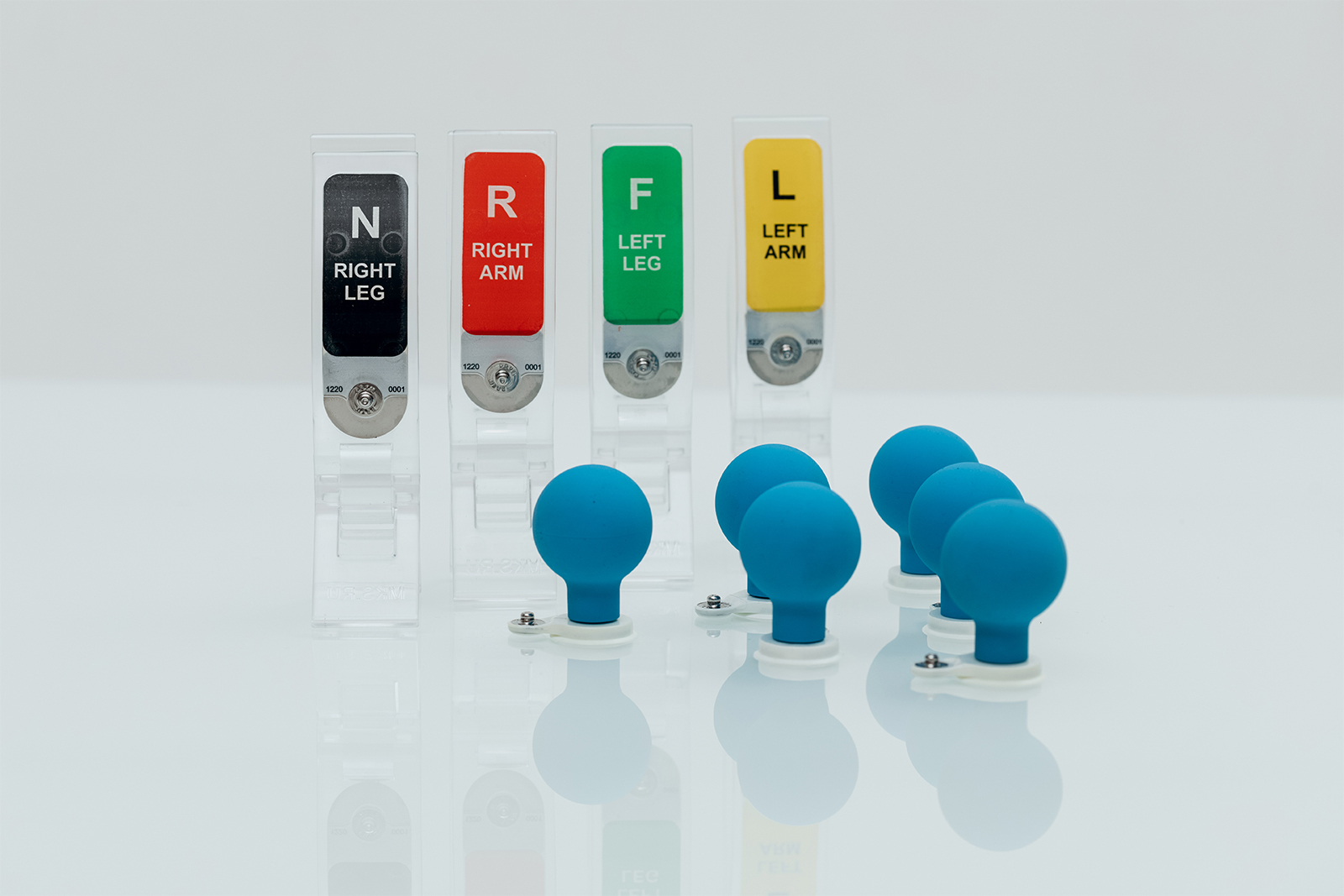
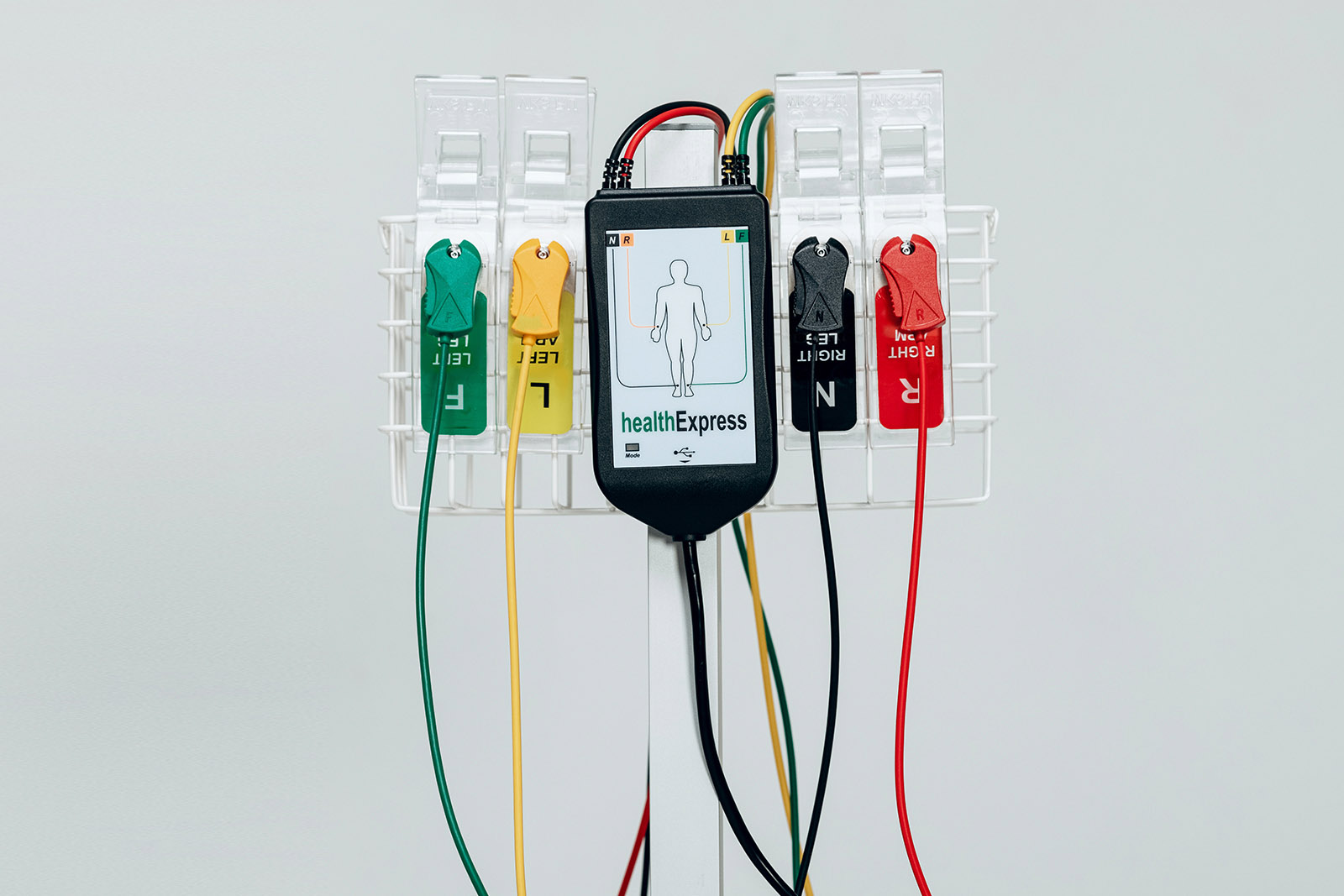
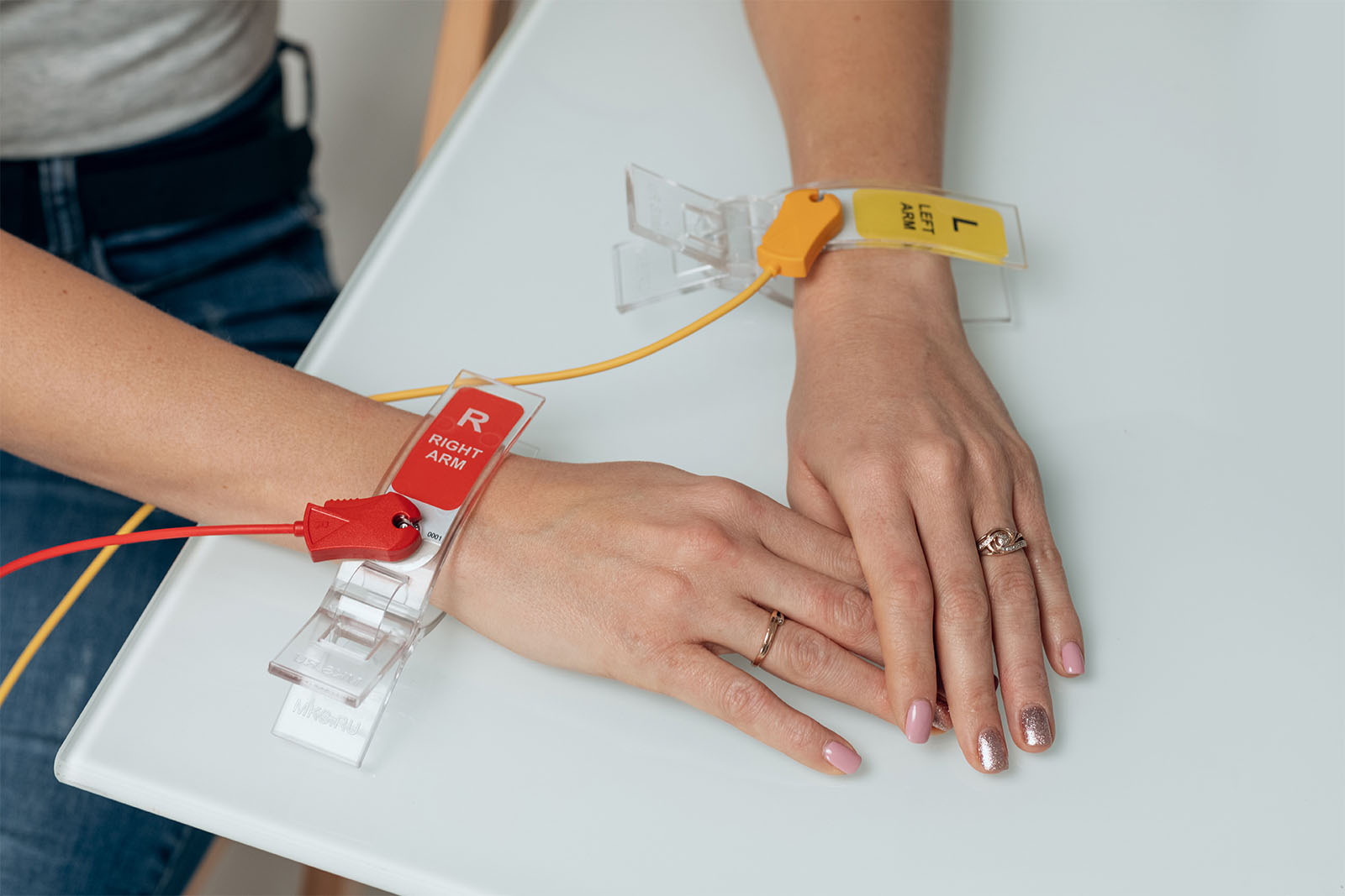
Utilizing 4 limb electrodes to deliver a highly exective heart health screening solution. Simple computer interpretation provides for increased patient-doctor.
interaction with the results delivered in just 3 minutes without the need to remove clothing. Compact and fully portable, the device requires only minimal training for ease of interpretation using the visualization.
HealthExpress uses two methods: heartVUE for measuring ECG microalternations, Heart Rate Variability analysis for determination of its functional state.
SOFTWARE
HealthExpress system was developed using the concept of modularity, which allows you to select the composition of the complex for specific tasks.
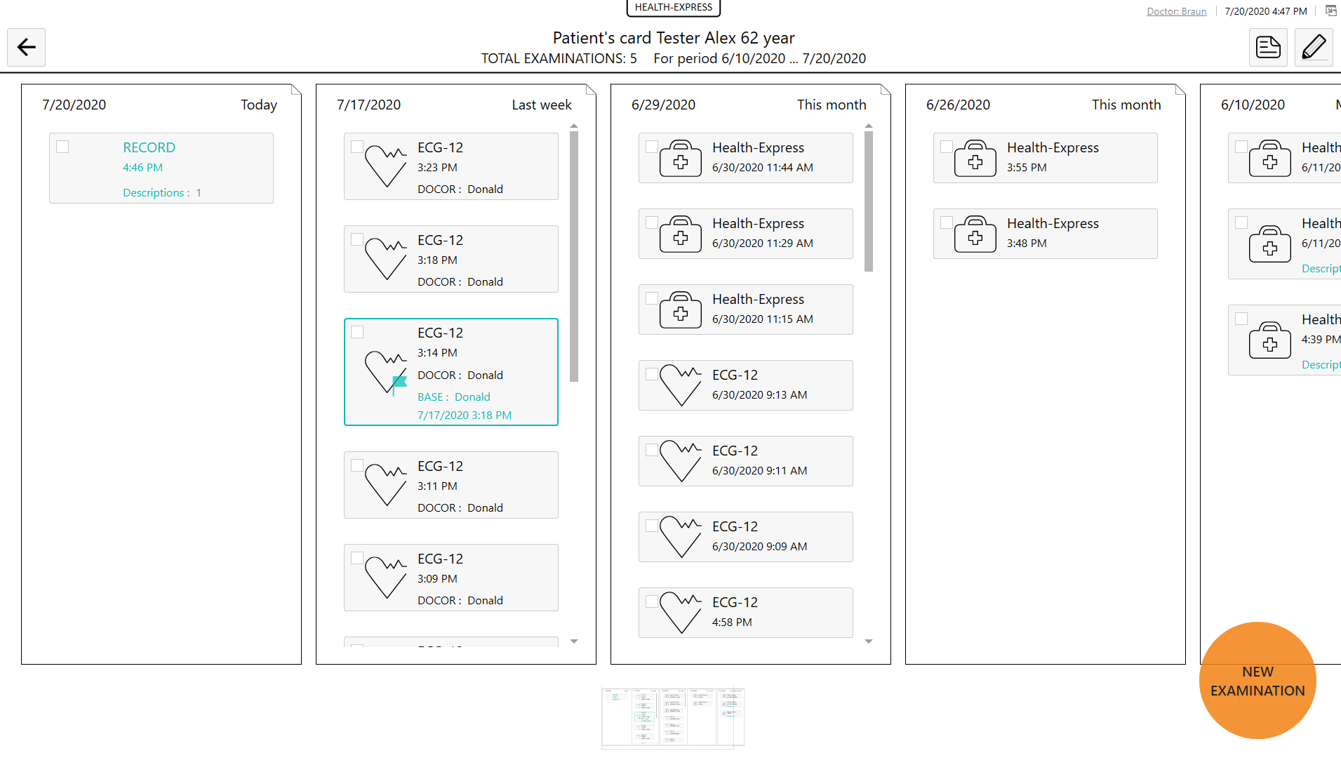
HealthExpress system modules are additional programs or hardware and software solutions that are connected to the patient database.The database provides organization and storage of data about patients and the results of their examinations, as well as communication with modules, on the one hand, and with external medical information systems (MIS) on the other side.

«CARDIOVISOR» (DISPERSIVE ECG MAPPING)
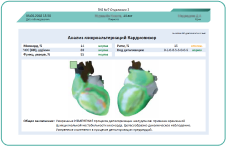
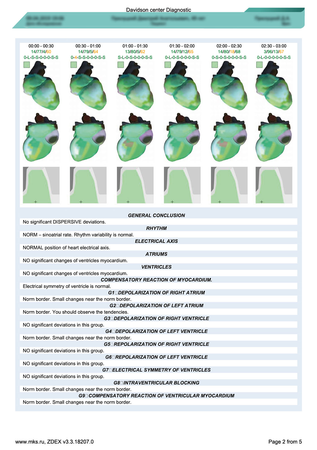
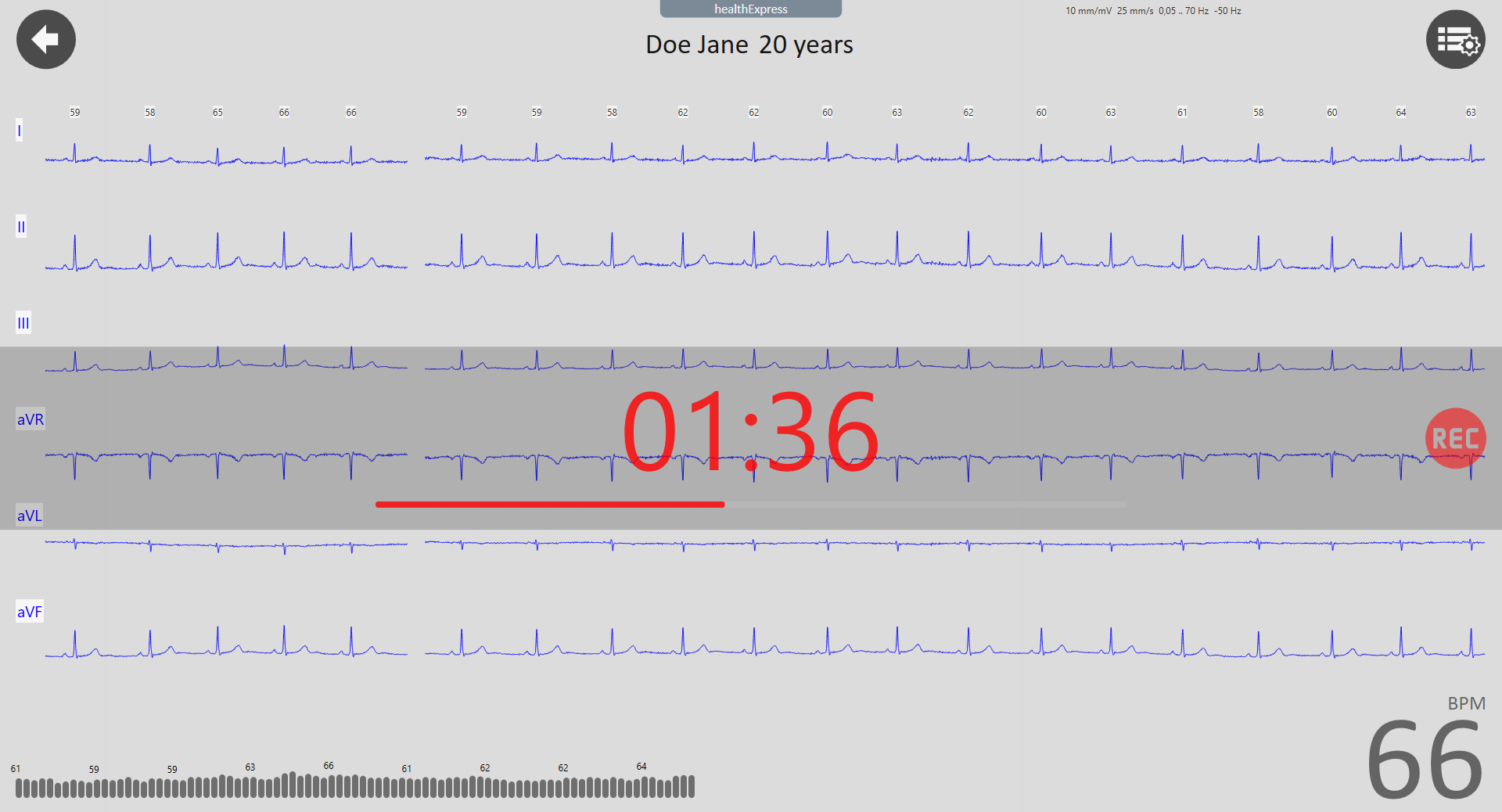
The module is designed for non-invasive screening assessment of the state of the heart and the level of stress in the body by analyzing ECG microalternations, the so-called. assessment using the “ECG dispersion mapping” method. During the examination, a resting ECG is recorded in a sitting or lying position from 4 electrodes placed on the limbs. The health worker selects the registration duration: 30 seconds, 3 or 5 minutes.
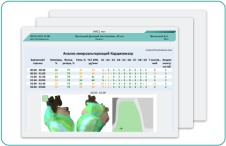
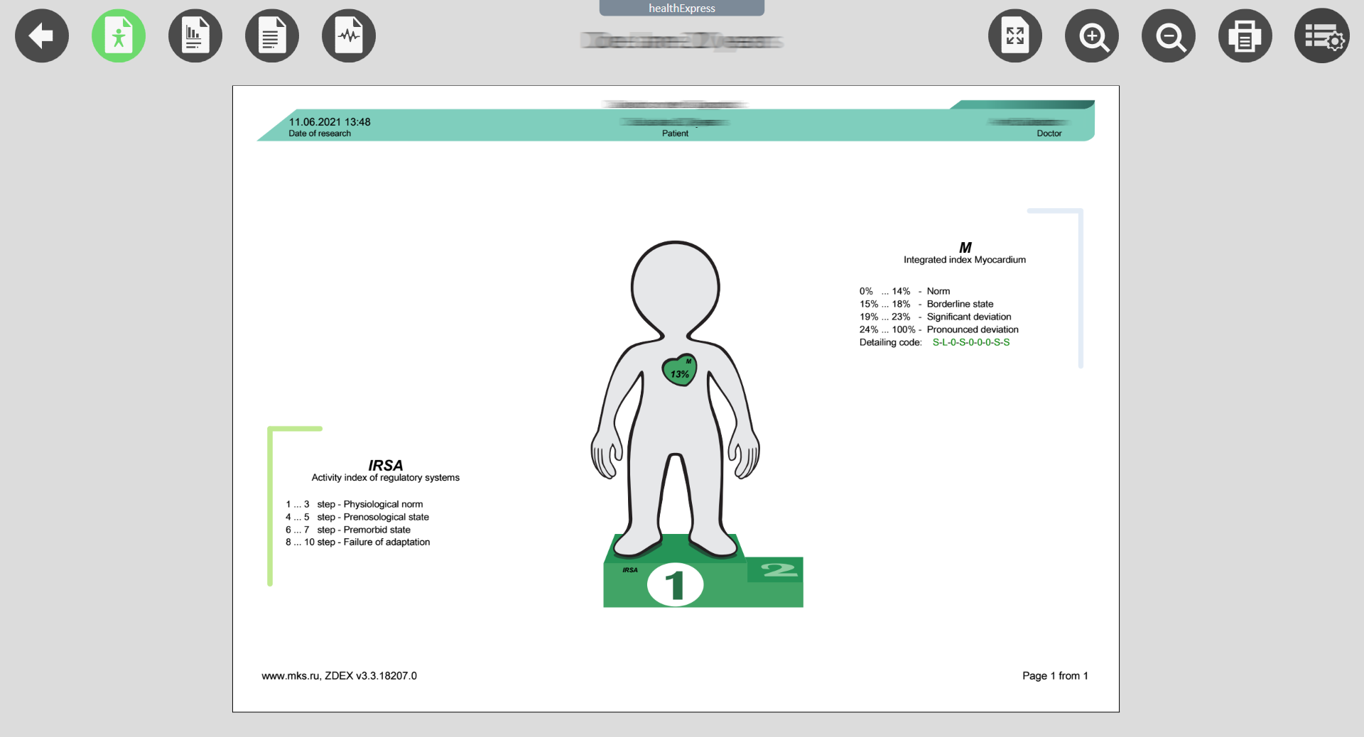
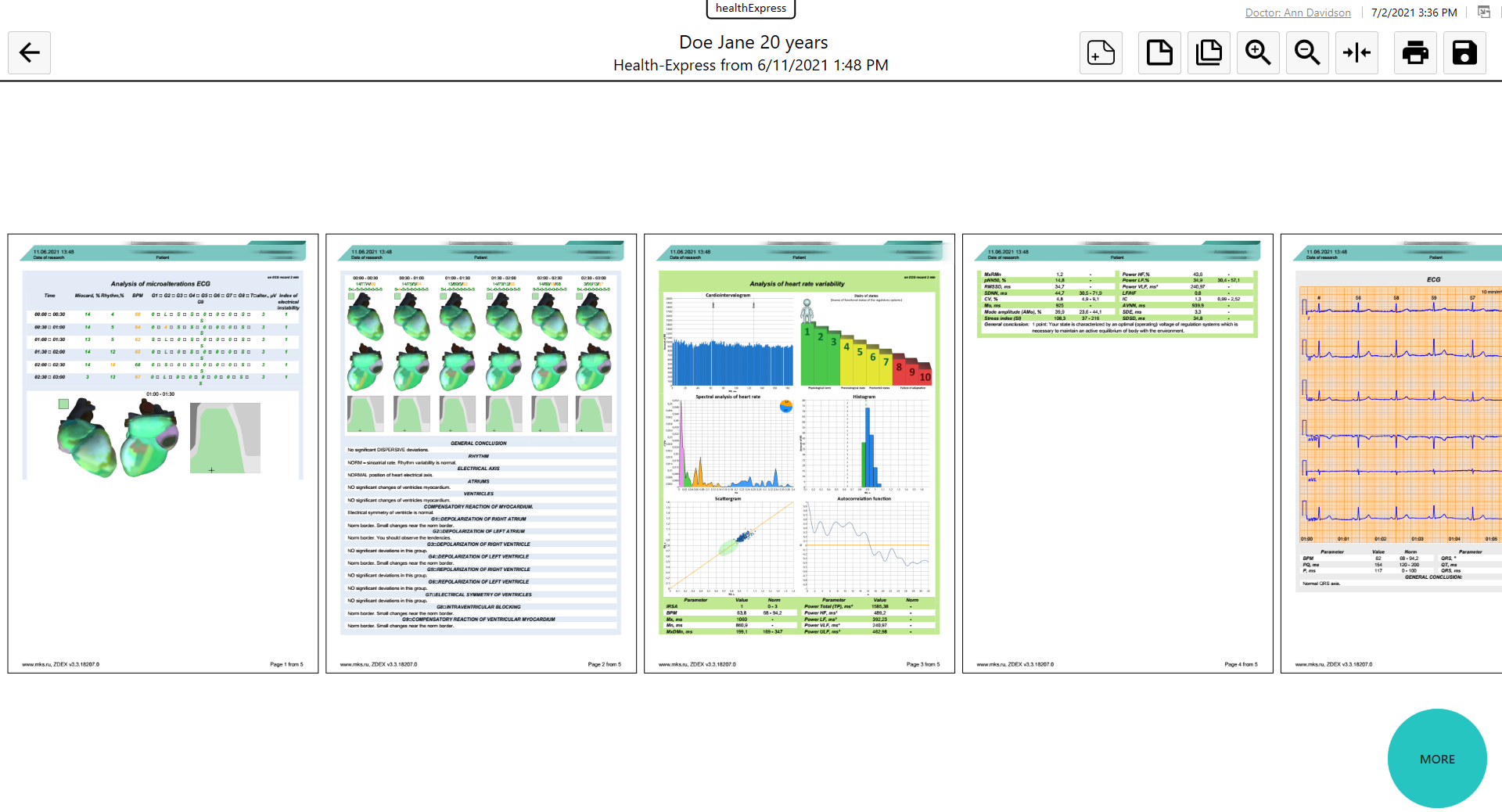
The characteristics of microalternations are represented by the integral index "MYOCARD"* and nine indicators detailing changes in the chambers of the heart, as well as in the de- and repolarization intervals. A map of averaged amplitudes of microalternations is formed in the form of a 3-dimensional color model of the heart (dispersion portrait of the heart).
The ECG microalternation analysis technique selects 30-second sections from the ECG recording. For each section, an automatic analysis of low-amplitude oscillations of the ECG signal is carried out in successive heart contractions, the so-called. analysis of microalternations. This is fundamentally different from standard ECG contour analysis. Microalternations are sensitive indicators of the overall functioning of the physiological systems of the body involved in the mechanisms of heart regulation. The DC EC technique responds to changes in the ion balance in myocytes, shifts in sympathoadrenal activation and other metabolic changes that, due to their small magnitude, do not appear in the morphology of the ECG or on ultrasound of the heart. Similarly, the DC ECG technique responds to the hidden dynamics of the compensatory reaction of the left ventricle, which makes it possible to timely identify the state of cardiac overload.

«ASSESSMENT OF HEART RATE VARIABILITY (HRV)»
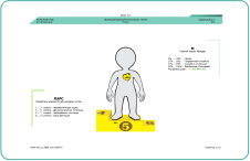
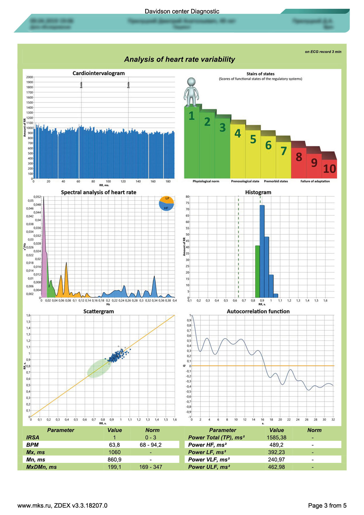
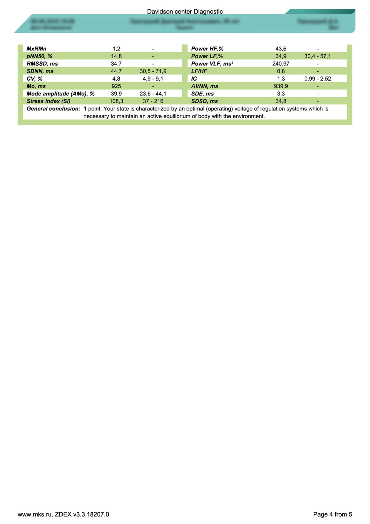
Analysis of heart rate variability (HRV) is a standard, scientifically based methodology for donosological diagnostics to obtain information about the degree of tension of regulatory systems, as a nonspecific response of the body to any adverse effects that require the mobilization of functional reserves. The method for assessing HRV using a 3- or 5-minute ECG recording calculates time and frequency parameters, and also classifies the functional state of the body based on the concept of homeostasis and adaptation with the calculation of the Activity Index of Regulatory Systems (PARS).

«ECG-12»
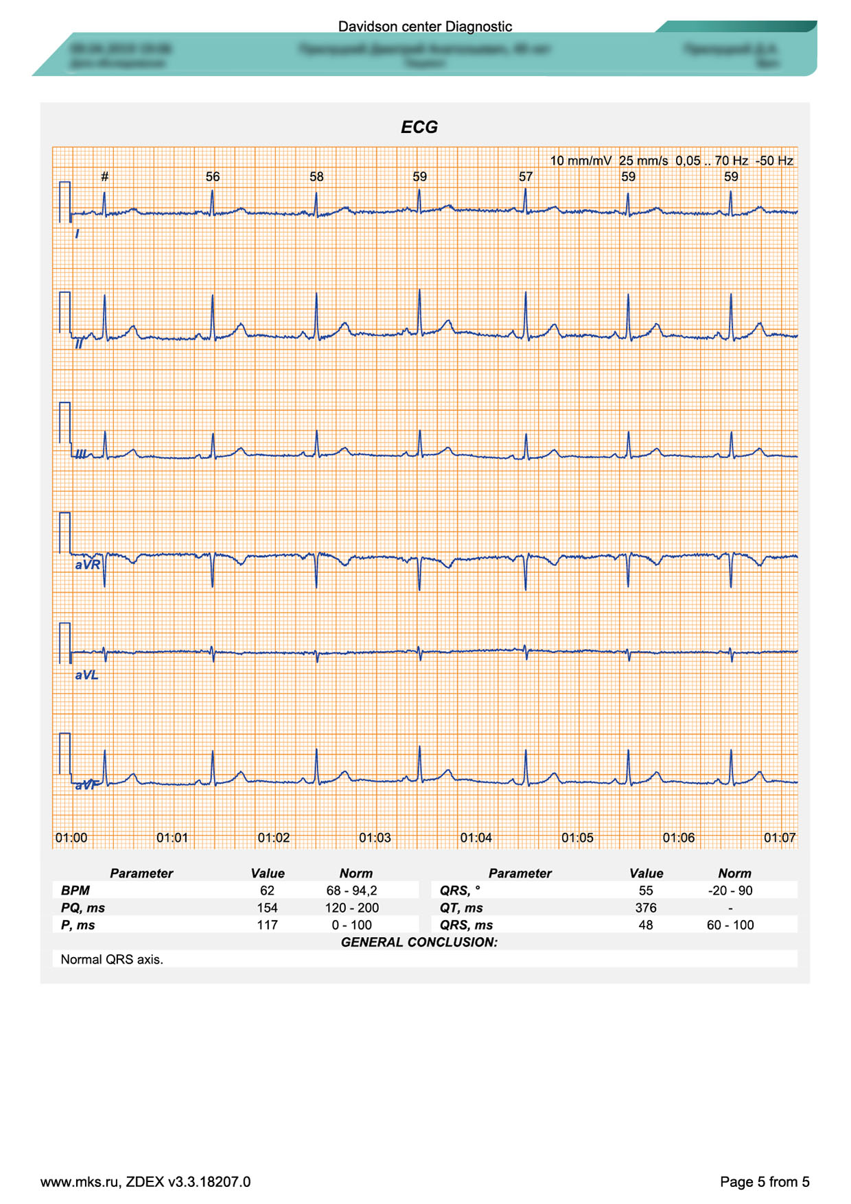
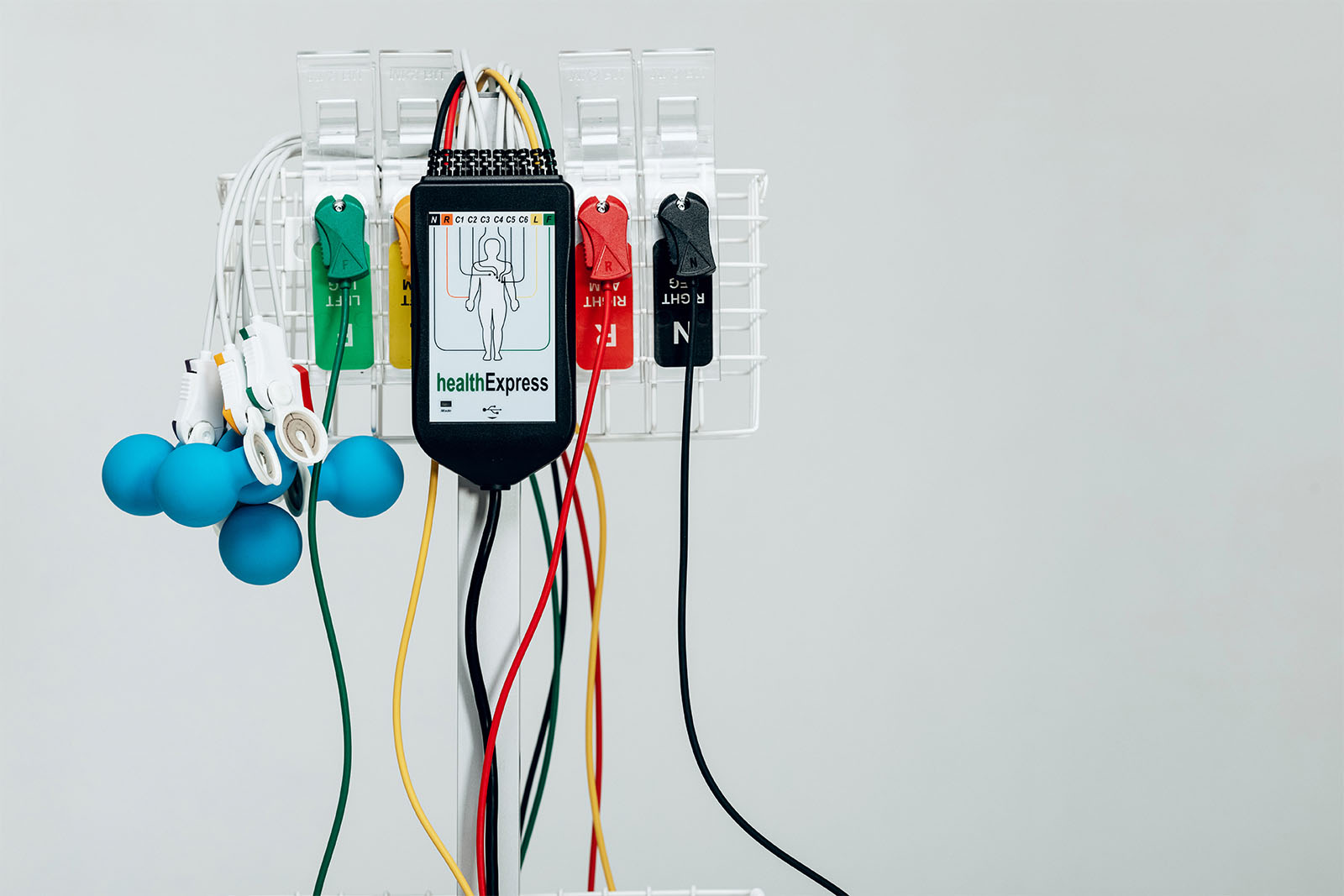
The ECG-12 module allows you to take a 12-channel electrocardiogram and carry out its contour analysis. A surface ECG is a display of the total vector of the electrical activity of the heart on the patient’s skin, which is recorded by an ECG amplifier as the difference in electrical potential between the electrodes. These potential differences are called ECG leads. The 12 standard lead scheme is widely used. Six of them are conventionally vertical, obtained from frontally located electrodes on the limbs (leads I, II, III, aVR, aVL, aVF) and six are conventionally horizontal, obtained from electrodes located on the chest in the precordial region (leads V1, V2, V3, V4, V5, and V6).
The 12-lead ECG is a decisive research method for establishing a large number of cardiac diagnoses, especially arrhythmias and myocardial ischemia. An ECG can also diagnose atrial enlargement, ventricular hypertrophy, and conditions that can lead to syncope or sudden death, such as Wolff–Parkinson–White (WPW) syndrome, long QT syndrome, and Brugada syndrome.
SPECIFICATION
| Parameter | Value |
| Electrical safety | class II type CF |
| Electromagnetic compatibility | group 1, class B as per CISPR 11 |
| Protection | IPX0 as per IEC 60529 |


