OVERVIEW
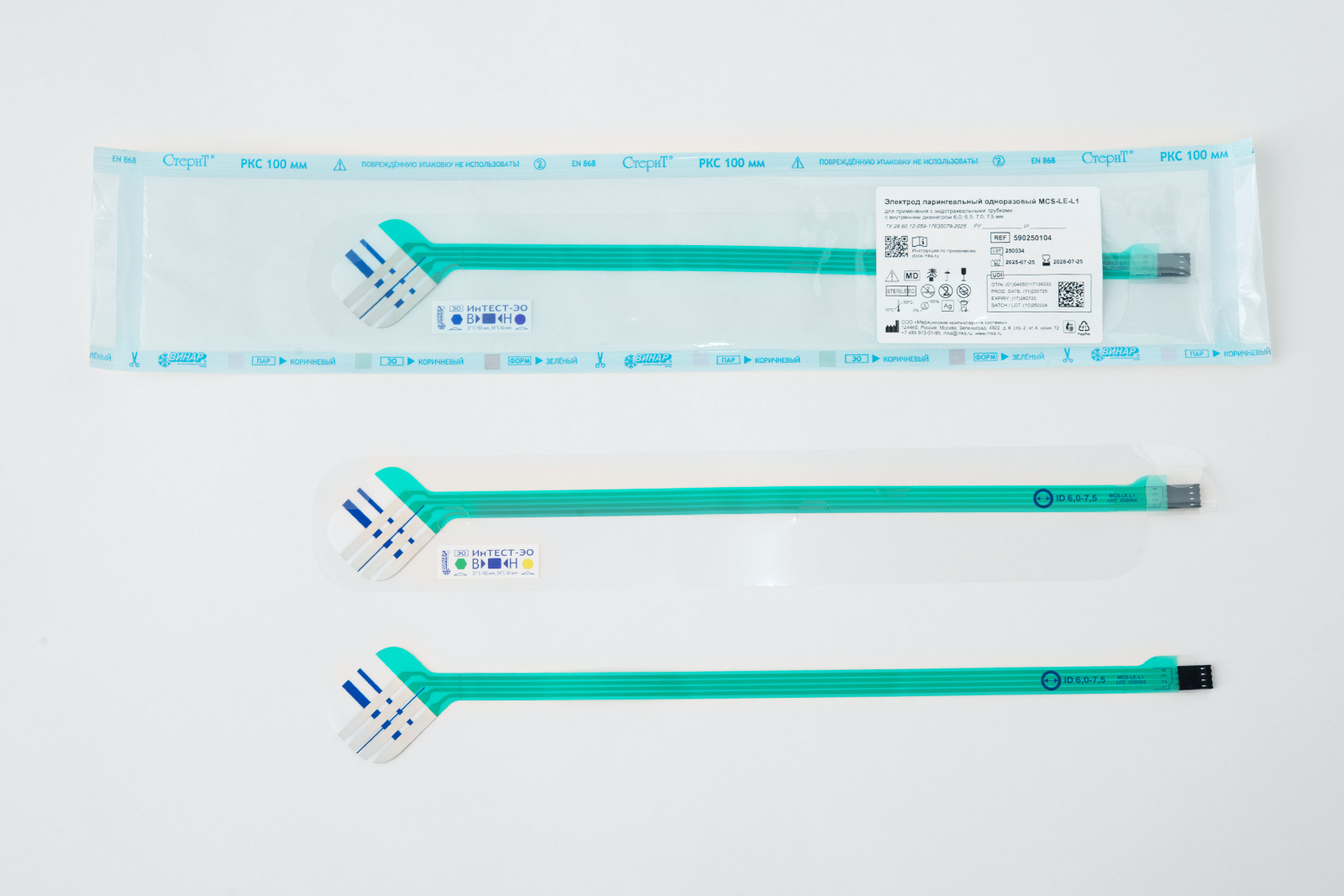
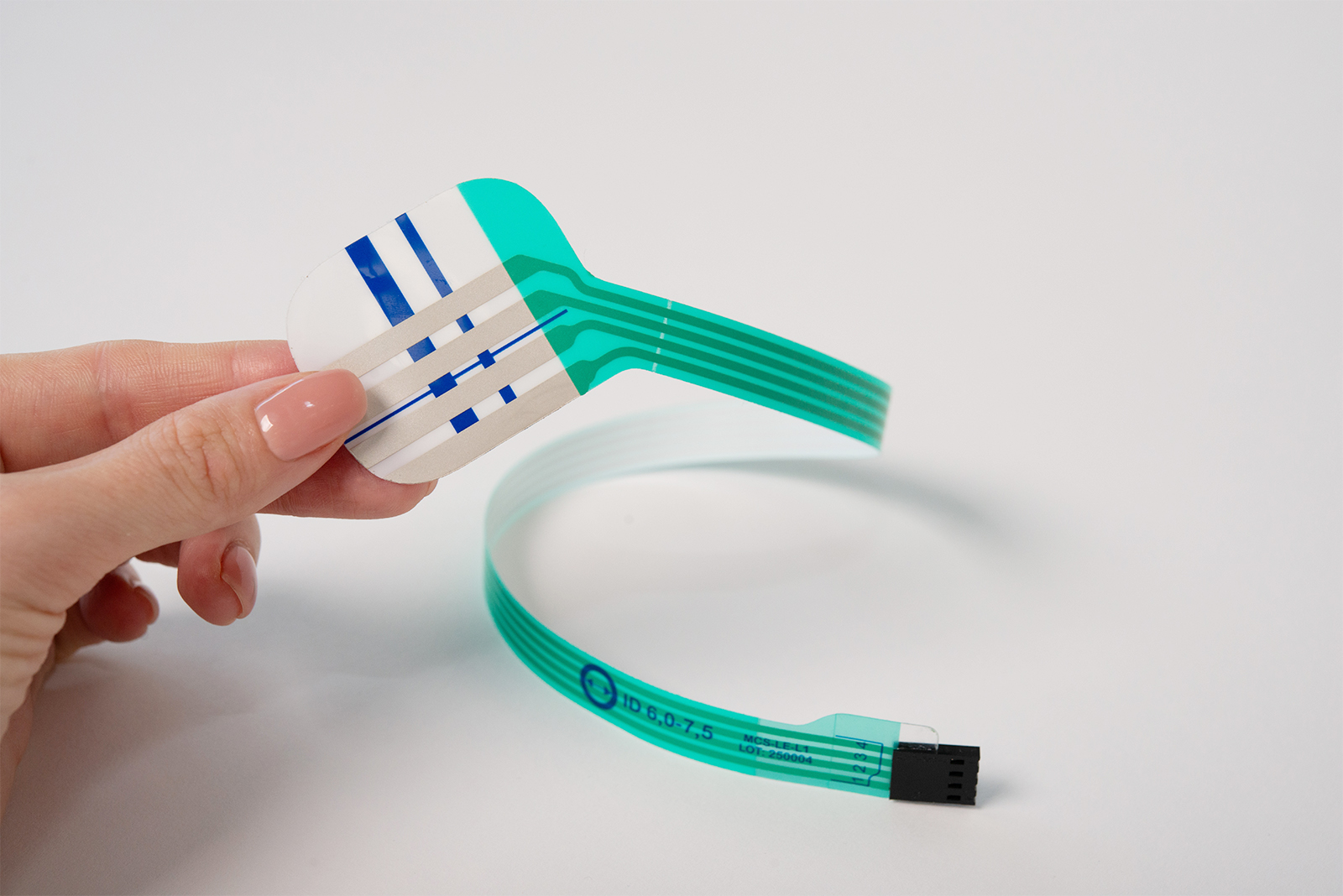
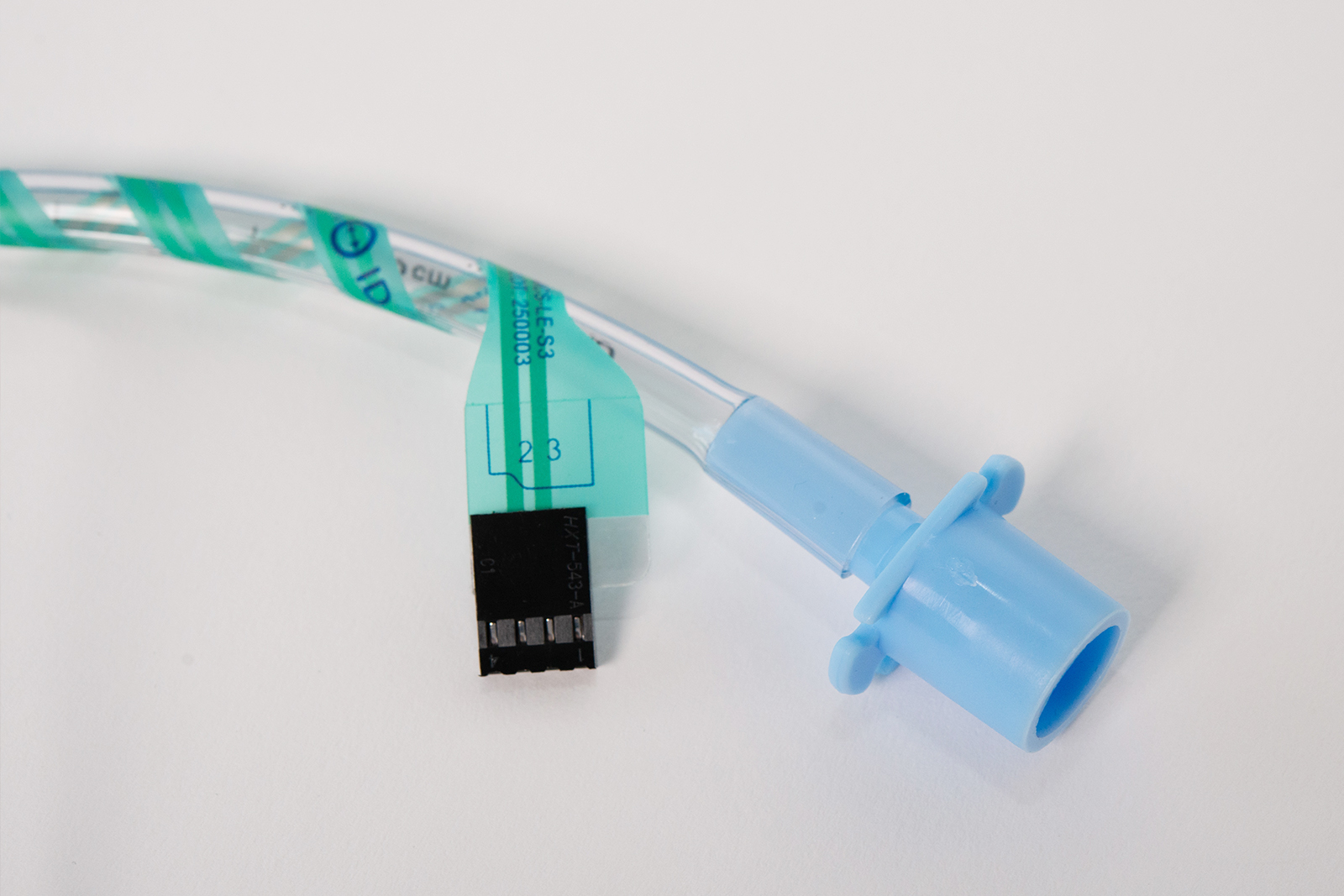
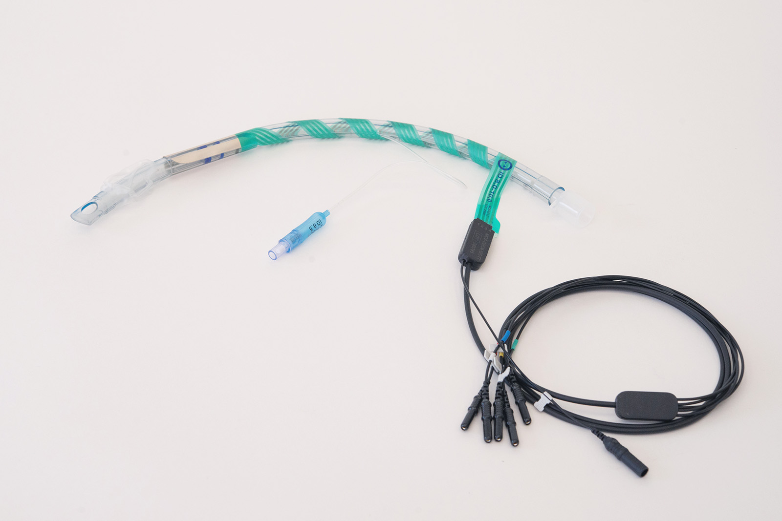
Disposable laryngeal electrode MCS-LE are designed for recording electromyographic (EMG) activity of the laryngeal muscles and vocal cords during surgical procedures.
The electrodes are recommended for use in operations with a high risk of unintentional damage to motor nerves and where visual identification is not possible.
The electrode is placed on an endotracheal tube and inserted together with it into the patient's larynx. The current-carrying part of the electrode contacts the vocal cords, allowing the recording of the signal passing through the nerves when surgical instruments come into contact with them.
The electrodes are manufactured in five versions, corresponding to different sizes of endotracheal tubes.
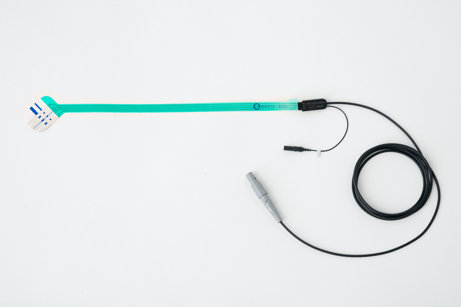
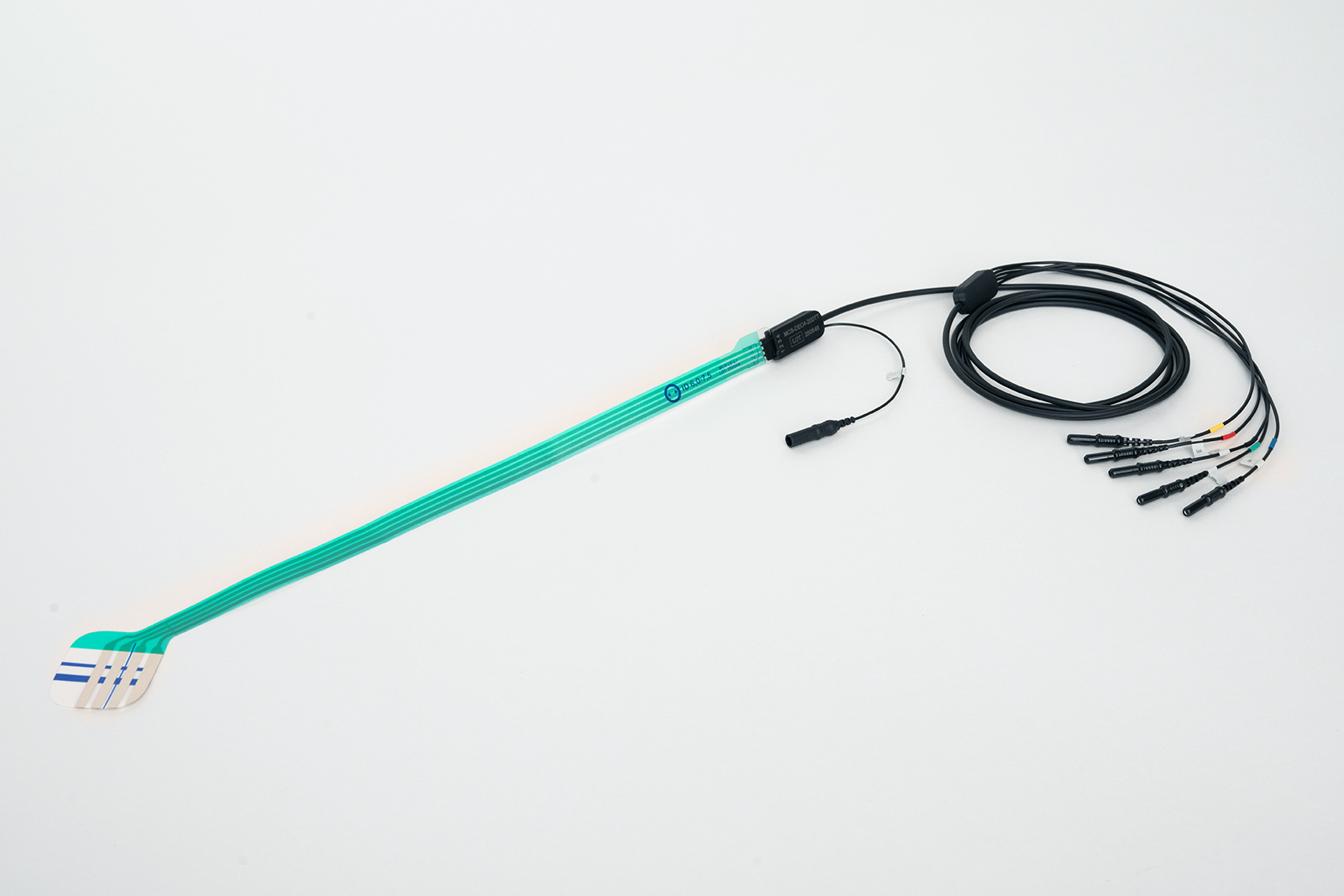
A dedicated reusable lead cable is used to connect the electrode to the recording device. The lead cable is available in several models, depending on the connector type and cable length.


SPECIFICATION
| Set up time | < 1 min |
| Time of continuous contact | > 24 h |
| The impedance of the electrode(at cutoff frequencies of 0 Hz and 104 Hz) | ≤ 100 Ω |
| Offset voltage | < 100 mV |
| Offset instability(in the passband of 0,05 Hz to 1 Hz, -3dB) | < 150 μV |
| Internal noise(in the range from 2 Hz to 104 Hz at -3 dB) | < 15 µV |
| Dielectric strength of electrodes insulation | > 500 V |
| Resistance of electrodes insulation | > 109 Ohm |
| Expiration date of electrode | 3 years |
| Service life of the lead cable | 1 year |
| Operating conditions | from +10 to +40 °С |
| Transportation conditions | from +10 to +30 °С, < 60 % RH at 25 °С |
AVAILABLE CONFIGURATIONS
| Modification | Compatible nominal internal diameter (ID) of the tube | Number of contacts recording the EMG signal | Number of bipolar channels |
| Disposable laryngeal electrode MCS-LE-S1 | 2.0; 2.5 mm | 2 | 1 |
| Disposable laryngeal electrode MCS-LE-S2 | 3.0; 3.5; 4.0 mm | 2 | 1 |
| Disposable laryngeal electrode MCS-LE-S3 | 4.5; 5.0; 5.5 mm | 2 | 1 |
| Disposable laryngeal electrode MCS-LE-L1 | 6.0; 6.5; 7.0; 7.5 mm | 4 | 2 |
| Disposable laryngeal electrode MCS-LE-L2 | 8.0; 8.5; 9.0; 9.5; 10.0 mm | 4 | 2 |


INSTRUCTIONS FOR USE
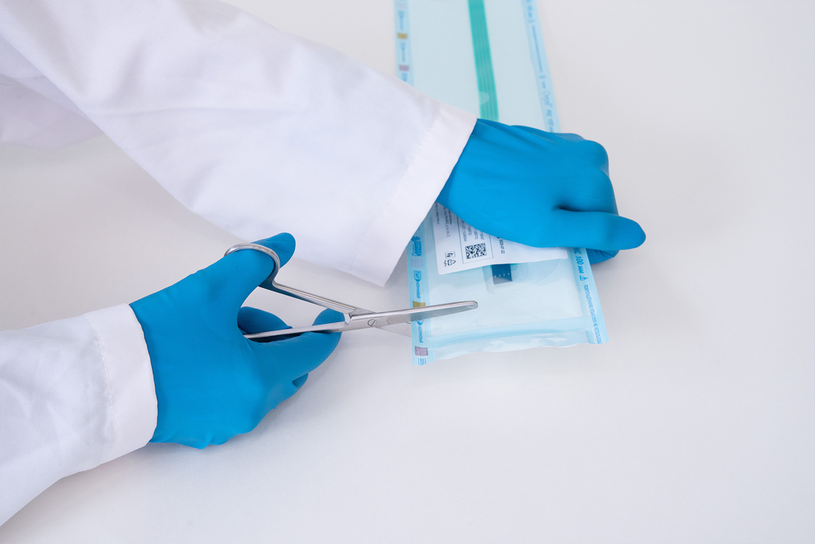
Step 1. Remove the electrode from its packaging. Verify that the selected electrode matches the nominal internal diameter (ID) of the endotracheal tube being used.
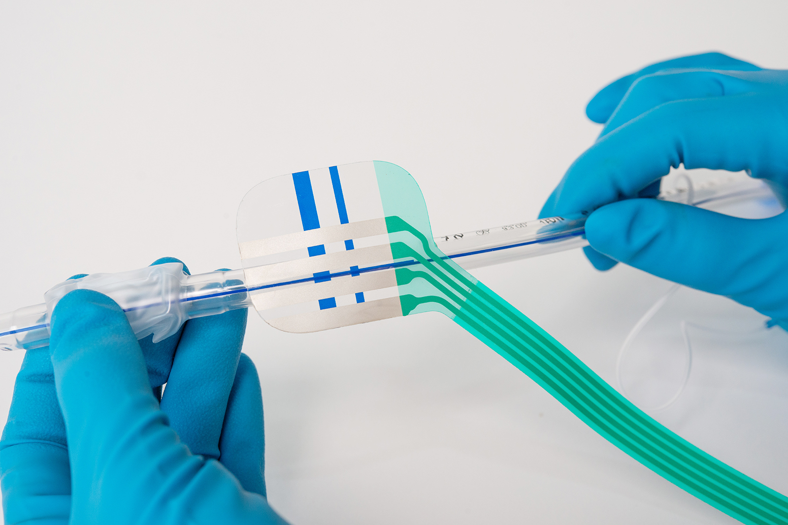
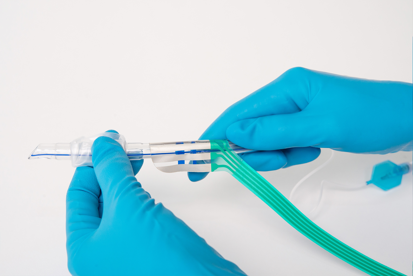
Step 2. Remove the protective backing from the adhesive layer of the contact portion of the electrode.
Step 3. Place the contact portion of the electrode onto the tube.
a. Align the depth marking on the electrode with the corresponding marking on the tube.
b. Orient the electrode along the longitudinal axis of the tube.
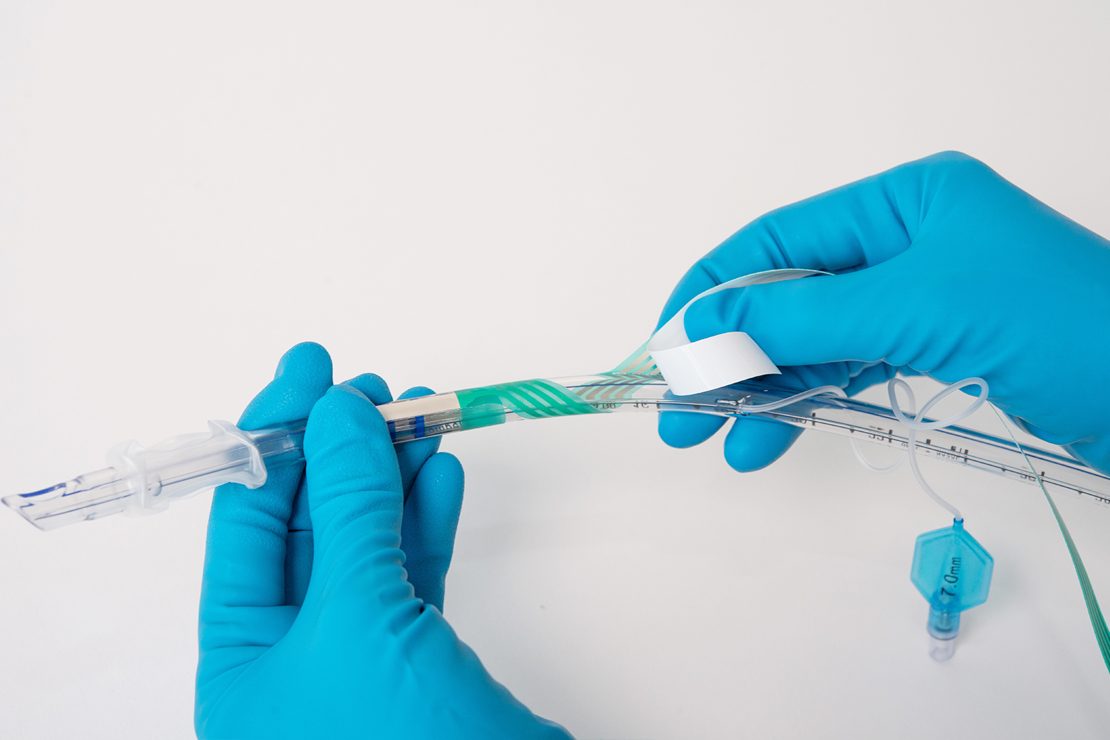
Step 4. Gradually apply the electrode to the tube while rotating it.
a. First, attach the larger part (with the non-conductive field).
b. Then, attach the smaller part, overlapping it onto the larger part.
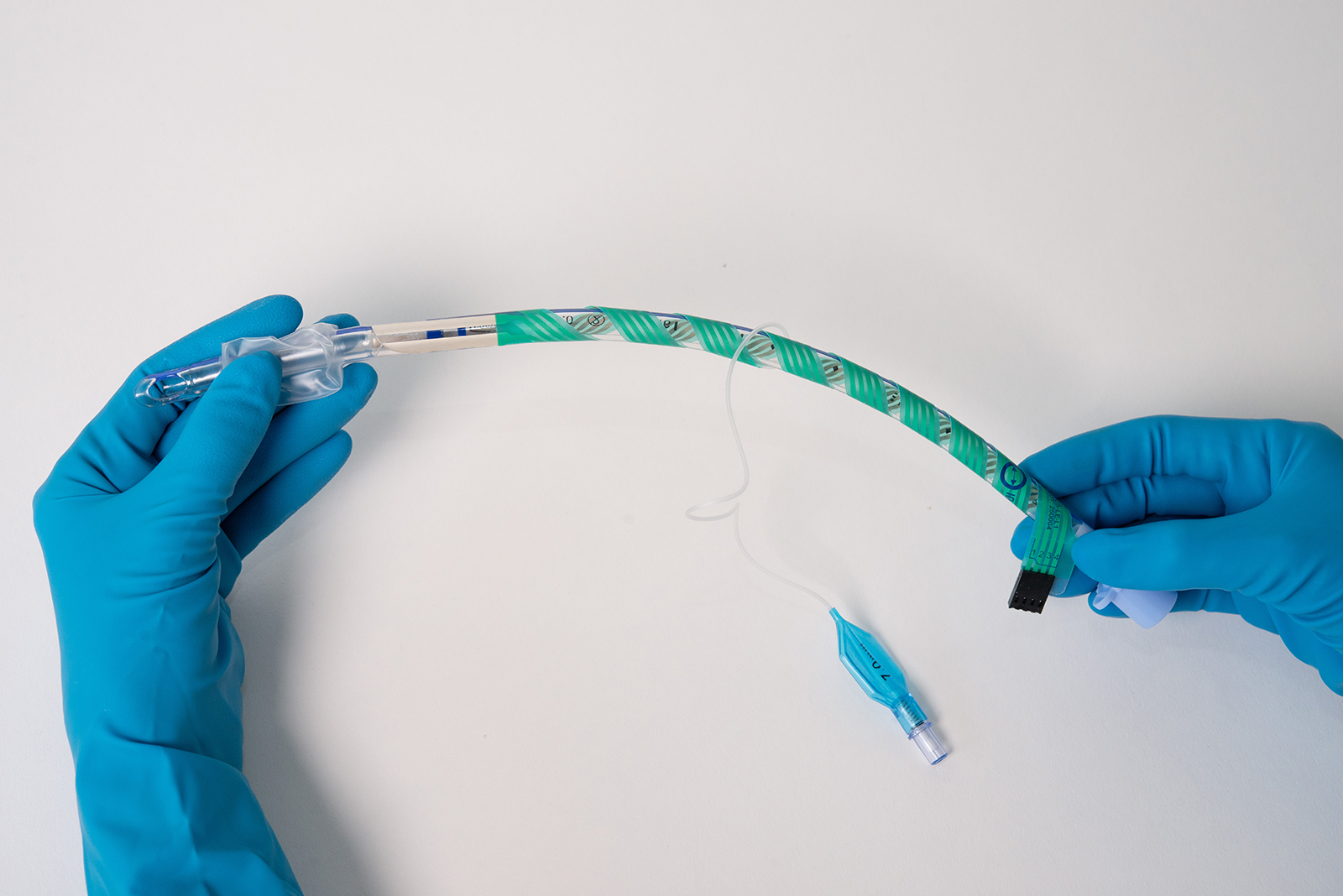
Step 5. Set the correct angle for wrapping the middle portion of the electrode (the lead/loop). Partially remove the protective backing from the adhesive layer of the loop. Do not allow the parts of the loop to stick to each other; straighten the tube and apply the electrode loop at an angle to it.
Step 6. Route the electrode connector to sit alongside the endotracheal tube connector. If necessary, the last turn of the loop around the tube may be additionally secured with adhesive tape to prevent accidental tension on the loop during connector attachment from causing it to peel off the tube or kink the loop.
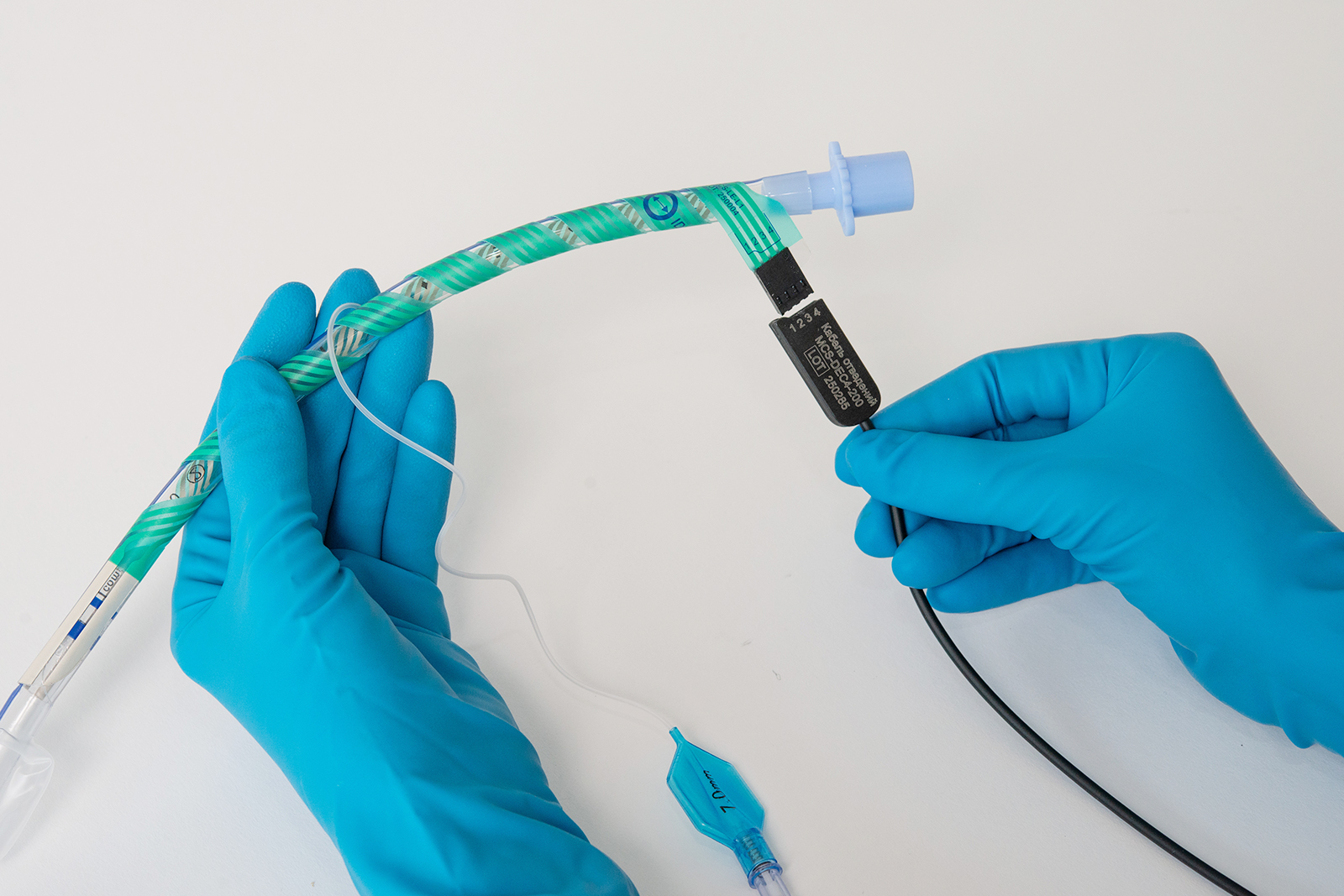
Step 7. Connect the reusable lead cable to the electrode connector.
Step 8. Connect the lead cable to the corresponding input jacks on the EMG recording device.

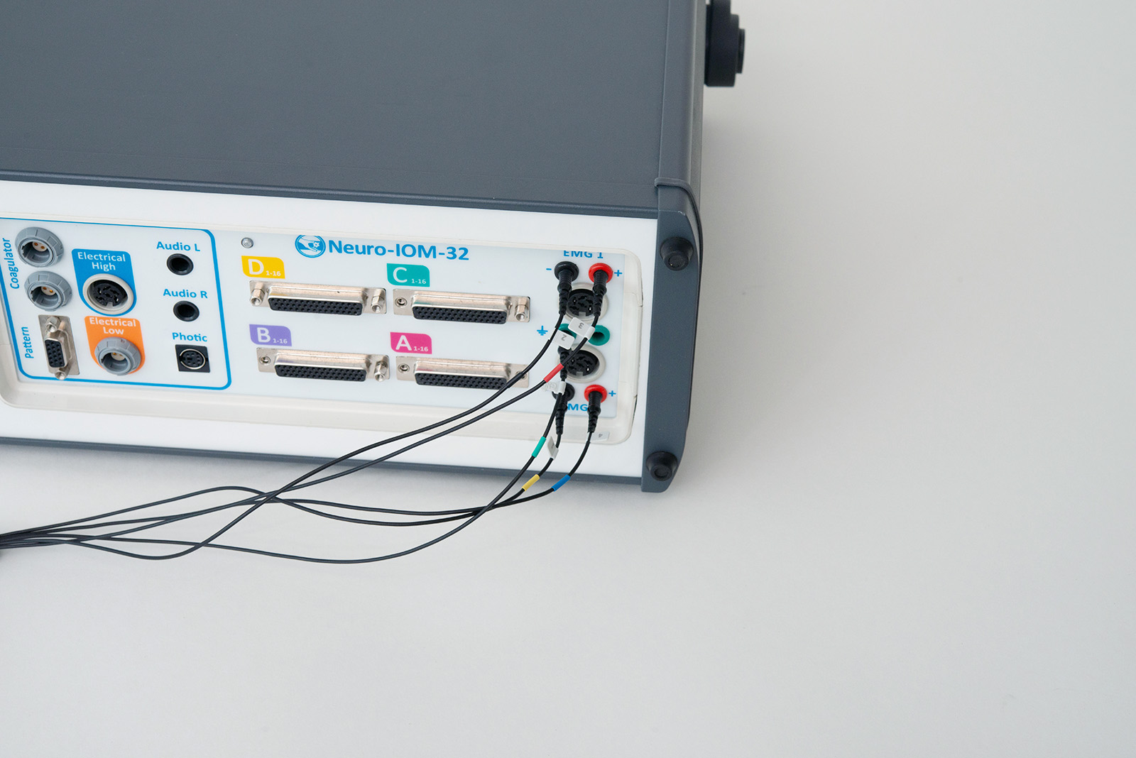
LEAD CABLES
| Model | Length | Connector Type |
| Lead Cable MCS-DEC4-120TT | 120 cm | 5 x TouchProof 1.5 mm (DIN 42802-ST) |
| Lead Cable MCS-DEC4-200TT | 200 cm | 5 x TouchProof 1.5 mm (DIN 42802-ST) |
| Lead Cable MCS-DEC4-200L0 | 200 cm | Redel male connector, 10-pole, 40° |
MCS-DEC4-120TT / 200TT
MCS-DEC4-200L0
PHOTO